Lumbar Sacral Disease

Lumbar Sacral Disease, Sacroiliac Disease
& Proximal Suspensory Desmitis

It has become evident over the past decade that when coming across sacroiliac problems there is a diverse range of treatment methods prescribed with a very mixed bag of success. In my experience, the importance of successful treatment is correct diagnosis.  For too long we have grouped a number of biomechanical attributes to ‘sacroiliac disease‘.  For the past 10 years, I have been trying to differentiate between lumbar sacral disease and sacroiliac disease because the success of the treatment and stabilising the condition long term depends on the structure involved.
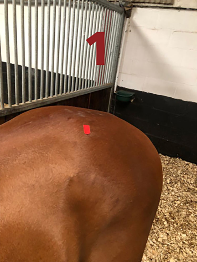
Sacroiliac

Picture 1 shows the sacroiliac joint deep underneath this point.
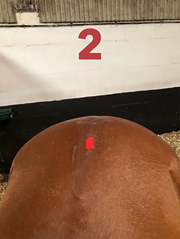
Sacroiliac

Picture 2 shows the lumbar sacral junction is deep underneath this point.
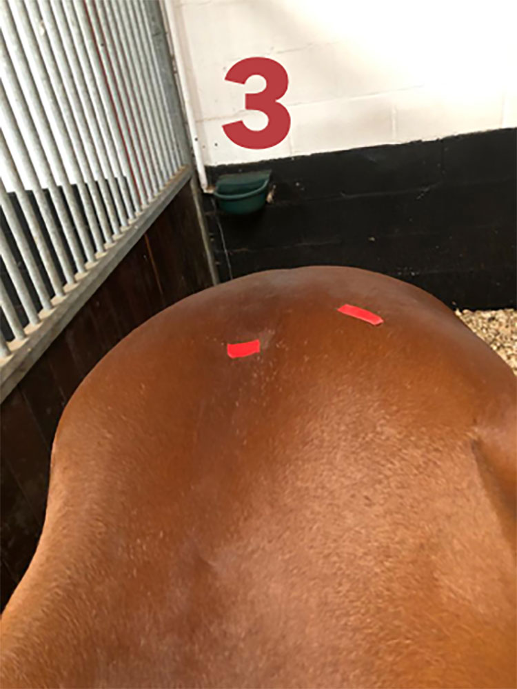
Sacroiliac

Picture 3 shows from the surface, the lumbar sacral region and sacroiliac area, which are deep underneath.


I produced a lecture as far back as 2010, linking sacroiliac disease with proximal suspensory ligament desmitis (PSLD).  I still firmly believe that the two are closely related and if both are targeted, then the success rate of the horse returning to function are greatly improved.  

No rehabilitation program is the same for every horse.  Each case MUST be taken individually.

However as a veterinary physiotherapist, my limitations are derived on the success of diagnosis by the veterinary surgeons.  I am lucky to work with some excellent vets that will try to send cases to be treated and managed for the horses well being and successful career to diagnose all areas of involvement in an abnormality. They will ultrasound scan or send horses for scintigraphy to diagnose accurately where a problem is initiating. (Lumbar sacral, sacroiliac or suspensory ligaments).

For the owners it is important to understand why we wish to differentiate between lumbar sacral and sacroiliac. The areas of treatment are anatomically very close, the distance between the lumbar sacral facet joint and the sacroiliac joint are actually  centimetres apart and therefore when corticosteroid is injected into the area it will diffuse to both areas  (the picture on the left clearly shows where the patient has been injected).

Sacroiliac

For the physio it is important, as the rehabilitation work program and lameness management of the condition is slightly different between Lumbar sacral disease and sacroiliac disease. All cases have numerous restrictions/ requirements to successful treatment.

This is why over the past 10 years I have specialised in rehabilitating cases at my premises.  Cases are far more successful when they have an intensive method of treatment in-house for a few weeks and then return home with a specific program for the individual and then they are enrolled on a lameness management program with myself and their veterinary surgeon.

Privacy Settings
We use cookies to enhance your experience while using our website. If you are using our Services via a browser you can restrict, block or remove cookies through your web browser settings. We also use content and scripts from third parties that may use tracking technologies. You can selectively provide your consent below to allow such third party embeds. For complete information about the cookies we use, data we collect and how we process them, please check our Privacy Policy
Youtube
Consent to display content from - Youtube
Vimeo
Consent to display content from - Vimeo
Google Maps
Consent to display content from - Google
Spotify
Consent to display content from - Spotify
Sound Cloud
Consent to display content from - Sound